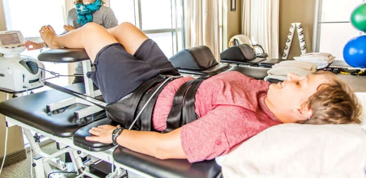
Explore effective chiropractic treatment options for carpal tunnel syndrome to alleviate pain and restore mobility.
Table of Contents
Abstract
This educational post explores the complexities of Carpal Tunnel Syndrome (CTS), a common yet often debilitating condition characterized by numbness, tingling, and pain in the hand and wrist. From my perspective as a practitioner with dual credentials in chiropractic (DC) and advanced practice nursing (FNP-BC), and certifications in functional medicine (CFMP, IFMCP), I will guide you through the anatomy, pathophysiology, and diagnosis of CTS. We will explore the latest findings from leading researchers, presenting a modern, evidence-based understanding of why this condition occurs and how it affects the body’s intricate neuromuscular system. This post will detail a comprehensive, integrative treatment model that combines conventional medical interventions, such as corticosteroid injections, with the foundational principles of chiropractic care, functional medicine, and targeted rehabilitation. A key focus will be our multidisciplinary clinical model at Injury Medical Clinic, P.A., where I work closely with our Medical Director, Dr. Maria Guadalupe Cardenas, MD, an experienced internist. Together, we blend medical oversight with chiropractic adjustments, soft tissue therapies, and lifestyle modifications to alleviate symptoms and address the root causes of CTS. I will walk you through a clinical procedure—a carpal tunnel injection—explaining the anatomical landmarks, technique, and rationale behind each step, and showing how these interventions fit within a broader, patient-centered care plan designed for lasting recovery.
As a clinician deeply invested in both the structural and functional aspects of human health, I regularly encounter patients struggling with Carpal Tunnel Syndrome (CTS). This condition, while localized to the wrist and hand, can profoundly affect a person’s quality of life, from their ability to work to simple daily tasks like buttoning a shirt or holding a coffee cup. At Injury Medical Clinic, P.A., in El Paso, Texas, we have cultivated a unique environment where multiple disciplines converge to provide comprehensive care.
Our approach is built on collaboration. I, Dr. Alex Jimenez, bring my expertise as a Doctor of Chiropractic (DC) and a Board-Certified Family Nurse Practitioner (FNP-BC), with advanced training in functional medicine. This dual perspective allows me to view conditions like CTS through biomechanical and systemic lenses. Crucially, Dr. Maria Guadalupe Cardenas, MD, provides medical direction for this care. Dr. Cardenas is a highly respected, Board-Certified Internist with over 40 years of clinical experience. As our Medical Director and Collaborative Physician (Texas MD License #J2933, NPI #1164426749), she is integral to our practice. This multidisciplinary structure ensures our patients benefit from rigorous medical oversight alongside holistic, non-invasive therapies. We integrate chiropractic care, advanced rehabilitation, functional medicine, and personal injury services to create a patient-centered treatment ecosystem. This lets us offer a spectrum of care, from conservative management to medically guided procedures, all under one roof.
Today, I want to take you on a journey into carpal tunnel syndrome, drawing on modern research and my clinical experience. We will explore what it is, why it happens, and how an integrative approach can offer a powerful path to healing. We will also walk through a specific clinical procedure—a carpal tunnel injection—to show how targeted medical interventions fit into our broader treatment philosophy.
Understanding the Carpal Tunnel: Anatomy of a Delicate Passageway
To truly grasp carpal tunnel syndrome, we must first understand the anatomy of the carpal tunnel itself. Imagine a narrow, rigid passageway in the wrist, almost like a tunnel running from the forearm into the hand. This tunnel isn’t just space; it is a bustling thoroughfare for critical structures that let your hand feel and move.
- The Floor and Walls: The foundation of this tunnel is formed by the carpal bones, the small, cube-like bones of the wrist, arranged in a C-shaped arch.
- The Roof: A strong, fibrous band of connective tissue called the transverse carpal ligament (also known as the flexor retinaculum) spans this bony arch. This ligament is incredibly tough and does not stretch easily, which is a key factor in the development of CTS.
What Travels Through the Tunnel?
The carpal tunnel is a tightly packed space. Nine flexor tendons and one very important nerve pass through it.
- Flexor Tendons: These are the long, cord-like structures that connect the muscles in your forearm to the bones in your fingers and thumb. When your forearm muscles contract, they pull on these tendons, causing your fingers and thumb to bend or “flex.” Nine of these tendons run through the carpal tunnel.
- The Median Nerve: This is the star player in the carpal tunnel syndrome story. The median nerve is one of the three major nerves of the forearm and hand. It originates from the brachial plexus in the neck and shoulder area, travels down the arm, and passes directly through the carpal tunnel alongside the flexor tendons.
The median nerve is responsible for two primary functions in the hand:
- Sensation: It provides feeling—touch, temperature, and pain—to the palm side of the thumb, the index finger, the middle finger, and half of the ring finger.
- Motor Function: It controls several small muscles at the base of the thumb, known as the thenar muscles. These muscles are crucial for an opposable thumb, allowing you to pinch and grasp objects effectively.
Because the carpal tunnel is a fixed, unyielding space, any condition that causes swelling, inflammation, or reduced volume within this tunnel can compress the delicate median nerve. This compression is the fundamental cause of carpal tunnel syndrome (Aroori & Spence, 2008).
The Pathophysiology of Carpal Tunnel Syndrome: Why Does It Happen?
Carpal tunnel syndrome is essentially a compression neuropathy, meaning it is a condition caused by direct pressure on a nerve. When the median nerve is compressed within the carpal tunnel, its function is impaired. This impairment occurs through a cascade of physiological events.
Initially, the compression restricts venous outflow from the nerve. Blood can still get into the nerve via the arteries, but it has trouble leaving through the veins. This causes fluid to back up, or edema, within the nerve and surrounding tissues. This swelling further increases the pressure inside the already crowded carpal tunnel, creating a vicious cycle. As the pressure continues to rise, it eventually becomes high enough to impede arterial inflow, starving the nerve of the oxygen and nutrients it needs to function properly.
This lack of blood flow, known as ischemia, is what triggers the initial symptoms of CTS. The nerve fibers responsible for sensation are particularly sensitive to ischemia. When they are deprived of oxygen, they begin to fire erratically, sending abnormal signals to the brain. We perceive these signals as:
- Paresthesia: The classic “pins and needles” sensation.
- Numbness: A loss of sensation.
- Tingling or buzzing: An electrical or vibratory feeling.
These symptoms are often worse at night for a few reasons. Many people sleep with their wrists flexed, which naturally increases the pressure in the carpal tunnel. Additionally, during the day, moving our hands and arms helps pump fluid out of the tissues. At night, when we are still, this fluid can accumulate in the wrist, exacerbating the compression. This is why patients often report waking up with numb hands and feeling the need to “shake them out” to restore sensation.
If the compression is severe or prolonged, it can lead to more permanent damage. The myelin sheath, the protective, insulating layer around nerve fibers, can begin to break down. This process, called demyelination, slows down the speed at which nerve signals can travel. In the most advanced cases, the nerve fibers themselves, the axons, can be damaged or die off, a process known as axonal degeneration. This can result in:
- Constant numbness: The sensory loss becomes permanent.
- Thenar muscle atrophy: The muscles at the base of the thumb begin to waste away because they lose nerve signals. This leads to profound weakness in grip and pinch strength, making fine motor tasks difficult.
- Chronic pain: The nerve becomes so irritated that it sends constant pain signals to the brain.
Risk Factors and Contributing Causes
While the direct cause of CTS is median nerve compression, several factors can contribute to this pressure. It is often not just one thing, but a combination of factors that leads to the condition.
- Anatomical Factors: Some individuals are born with a smaller carpal tunnel, making them more susceptible to compression. Wrist fractures or dislocations can also alter carpal tunnel anatomy and lead to CTS.
- Repetitive Hand and Wrist Use: Occupations or hobbies that involve prolonged or repetitive wrist flexion and extension can contribute to CTS. This includes tasks like typing, using a mouse, working on an assembly line, or using vibrating power tools. These repetitive motions can cause inflammation and swelling of the flexor tendons, which then take up more space in the tunnel and compress the median nerve (Sevy & Varacallo, 2024).
- Systemic and Metabolic Conditions: Many underlying health issues can increase CTS risk by causing systemic inflammation or fluid retention. These include:
-
- Diabetes Mellitus: High blood sugar levels can damage nerves directly (a condition known as diabetic neuropathy) and also contribute to inflammation.
- Rheumatoid Arthritis: This autoimmune condition causes chronic inflammation of the joints and the synovial sheaths that surround the tendons, leading to swelling within the carpal tunnel.
- Hypothyroidism: An underactive thyroid gland can cause widespread fluid retention (myxedema), including in the carpal tunnel tissues.
- Obesity: Higher body weight is strongly associated with increased CTS risk, likely because it increases pressure within the tunnel and raises baseline systemic inflammation.
- Hormonal Changes: Fluid retention associated with pregnancy and menopause can temporarily increase pressure in the carpal tunnel, leading to symptoms. In pregnancy, these symptoms often resolve after delivery.
From a functional medicine perspective, we also look deeper into these systemic drivers. We investigate sources of chronic inflammation, such as gut dysbiosis, food sensitivities, or exposure to environmental toxins. We assess for nutrient deficiencies, particularly B vitamins (like B6 and B12), which are crucial for nerve health. Addressing these underlying systemic imbalances is a core tenet of our integrative approach, as it helps reduce the body’s overall inflammatory burden and can powerfully support local treatments.
The Role of Integrative Chiropractic Care in Managing CTS
As a chiropractor, my primary focus is on the body’s structure and its relationship to nervous system function. For carpal tunnel syndrome, this perspective is invaluable. While the problem manifests in the wrist, the median nerve’s journey begins in the neck. Therefore, a comprehensive chiropractic evaluation for CTS does not just look at the hand and wrist; it assesses the entire kinetic chain from the cervical spine to the fingertips.
The Concept of “Double Crush Syndrome”
A crucial concept in understanding the chiropractic approach to CTS is the “Double Crush Syndrome.” This theory, first proposed by Upton and McComas (1973), suggests that a nerve that is already compressed at one location (for example, in the neck) becomes more susceptible to injury and dysfunction at another location further down its path (such as the wrist).
Think of it like a garden hose. If you have a small kink in the hose near the spigot, the water flow is already slightly reduced. If you then step on the hose further down the line, the second compression will have a much greater effect than if the hose had been perfectly straight to begin with.
In the context of CTS, the median nerve’s roots exit the spinal cord through small openings between the cervical vertebrae called neural foramina. If these vertebrae are misaligned (subluxation), or if there is a bulging or herniated disc or arthritic changes in the neck, these nerve roots can become irritated or compressed. This “first crush” in the neck may not be severe enough to cause symptoms on its own. Still, it can impair the normal physiological transport of nutrients and signaling molecules along the nerve axon (a process called axoplasmic flow). This makes the nerve metabolically compromised and more vulnerable. When this already-vulnerable nerve then passes through the carpal tunnel and encounters a “second crush” due to inflammation or repetitive strain, the symptoms of CTS are more likely to develop and can be more severe.
Our Chiropractic and Rehabilitative Approach
Our integrative treatment plan for CTS is therefore multifaceted and addresses all potential points of nerve entrapment and dysfunction.
- Cervical Spine Adjustments: Using precise, gentle adjustments, we work to restore proper motion and alignment of the neck vertebrae. The goal is to relieve any direct or indirect pressure on the median nerve roots, improving nerve signaling and axoplasmic flow from the origin point. This addresses the “first crush.”
- Extremity Adjustments: We also perform specific adjustments to the wrist (carpals), elbow, and shoulder. Misalignments in these joints can alter the biomechanics of the entire arm, contributing to wrist strain. For example, mobilizing a restricted carpal bone can sometimes help increase space within the carpal tunnel.
- Soft Tissue Therapies: The muscles and fascia of the forearm play a huge role in CTS. Overworked flexor muscles can become tight, shortened, and develop trigger points, which can contribute to tendon inflammation. We use a variety of techniques to address this:
-
- Myofascial Release: This involves applying sustained pressure to the fascial connective tissue to release restrictions and improve tissue mobility.
- Active Release Technique (ART): A movement-based massage technique that treats problems with muscles, tendons, ligaments, fascia, and nerves.
- Graston Technique: This uses specially designed stainless-steel instruments to detect and treat scar tissue or adhesions in muscles and tendons.
- Targeted Rehabilitation and Ergonomics: We teach patients specific nerve and tendon gliding exercises. These gentle movements encourage the median nerve and flexor tendons to slide smoothly past one another within the carpal tunnel, helping break up minor adhesions and improve mobility (Bland, 2005). We also provide crucial education on ergonomics—modifying the workstation and daily activities to reduce wrist strain. This can include adjusting chair height, keyboard position, and using ergonomic mice or wrist supports.
- Functional Medicine Support: As mentioned earlier, we use our functional medicine expertise to identify and address systemic contributors. This may involve dietary changes to reduce inflammation, targeted supplementation with nerve-supportive nutrients like alpha-lipoic acid and B vitamins, and lifestyle coaching to manage stress and improve sleep.
This comprehensive chiropractic and rehabilitative framework is designed to address the root biomechanical and physiological causes of the nerve compression, not just mask the symptoms. It is a foundational part of our integrative model.
Integrating Medical Interventions: A Clinical Walkthrough of a Carpal Tunnel Injection
In some cases, particularly when inflammation is significant and symptoms are acute, conservative care alone may not provide rapid relief. This is where integrating medical interventions, under Dr. Cardenas’s careful oversight, becomes so valuable. One of the most common and effective medical treatments for CTS is a corticosteroid injection. The goal is to deliver a powerful anti-inflammatory medication directly to the site of the problem—the carpal tunnel—to quickly reduce swelling and relieve pressure on the median nerve.
Let me walk you through how we perform this procedure, just as I did recently with a 65-year-old patient presenting with classic CTS symptoms in her right hand. My dual training as a nurse practitioner and a chiropractor allows me to bridge the gap between these two worlds, ensuring the procedure is performed with anatomical precision and within the context of a holistic treatment plan.
Step 1: Identifying the Anatomical Landmarks
Precision is paramount in any injection procedure. The goal is to deliver the medication to the target tissue while avoiding injury to surrounding structures, especially the median nerve itself. To do this, we must first meticulously identify our anatomical landmarks on the patient’s wrist.
- The Distal Palmar Crease: This is the most prominent crease across the palm, at the base of the hand where it meets the wrist. This crease serves as our primary horizontal reference, as it roughly overlies the entrance to the carpal tunnel. I begin by marking this line.
- The Palmaris Longus Tendon: This slender tendon runs down the center of the forearm and crosses the wrist. Interestingly, about 15% of the population is born without this tendon. To find it, I ask the patient to flex their wrist and touch their thumb and little finger together. If present, this action will cause the tendon to “pop out” in the middle of the wrist. In my recent patient, her palmaris longus was present but diminutive, meaning it was small and not easily visible, though I could feel it with palpation. The median nerve typically lies just deep and slightly to the radial side (thumb side) of the palmaris longus tendon.
- The Flexor Carpi Radialis (FCR) Tendon: This is another major tendon located on the thumb side of the wrist. To identify it, I ask the patient to make a fist and flex their wrist. The FCR tendon becomes firm and palpable. I can feel it running just to the side of where I located the palmaris longus.
By identifying these two tendons—the palmaris longus and the flexor carpi radialis—I have created a precise map of the underlying anatomy. The median nerve lies in the valley between them.
Step 2: Selecting the Injection Site and Approach
Several accepted approaches exist for a carpal tunnel injection. Clinicians often choose an approach based on preference and the patient’s specific anatomy. My preferred method is the flexor carpi radialis approach, as it provides a safe and reliable path to the carpal tunnel while minimizing the risk of directly hitting the median nerve.
- The Insertion Point: I choose an insertion point about 1 cm proximal to the distal palmar crease. “Proximal” in this context means closer to the elbow. This places the injection just before the tightest part of the carpal tunnel, allowing the medication to flow down into the tunnel.
- The Trajectory: My insertion point is located on the ulnar border of the flexor carpi radialis tendon (the side closer to the little finger). I aim the needle at a 30-degree angle, directed distally (toward the fingers) and slightly inward, targeting the space just deep to where the palmaris longus tendon would cross the distal palmar crease.
I mark both the insertion point and the target point with the retracted tip of a ballpoint pen. This creates a small indentation in the skin that serves as a visual guide without using ink that would be washed away during sterilization.
Step 3: Sterilization and Anesthesia
Infection control is non-negotiable. The injection site must be thoroughly cleaned to prevent introducing bacteria into the wrist.
- I first scrub the area with an alcohol pad, which also removes any ink lines I may have drawn.
- I then applypovidone-iodine (Betadine), a powerful antiseptic, twice. I apply it in a circular motion, moving from the center of the injection site outward.
To make the procedure as comfortable as possible, I use a topical vasoconstrictor spray, such as EMLA cream. This spray rapidly cools the skin, providing a brief but effective numbing effect. I spray it directly on the insertion site and wait for the skin to “flash white,” indicating it is cold enough to be numb.
Step 4: Preparing the Injection
For this procedure, I prepare a syringe with a combination of two medications:
- 1 mL of 1% Lidocaine (without epinephrine): This local anesthetic provides immediate pain relief. We use lidocaine without epinephrine because epinephrine is a vasoconstrictor (it narrows blood vessels). Lidocaine should. Lidocaine is generally avoided in injections around nerves and in areas with limited circulation, like the fingers.
- 1 mL of a Corticosteroid Solution: This is the therapeutic agent. Corticosteroids (such as triamcinolone or methylprednisolone) are powerful anti-inflammatory drugs. When injected into the carpal tunnel, the steroid reduces inflammation and swelling of the synovial sheaths surrounding the flexor tendons. This reduction in swelling increases space within the tunnel, relieving pressure on the median nerve (Marshall et al., 2007).
I use a 25-gauge, 1-inch needle. The 25-gauge needle is relatively fine, which helps minimize patient discomfort. The 1-inch length is sufficient to reach the target depth within the carpal tunnel.
Step 5: The Injection Procedure
With the patient’s hand resting comfortably, I give them clear instructions. This is critical to patient safety and cooperation.
“I am about to begin. The goal is to place the medicine around the nerve, not in it. If at any point you feel a sharp, electrical ‘zing’ or tingling that shoots into your fingers, that’s a sign that the needle tip is very close to or touching the nerve. You mustn’t jerk your arm away. Just stay calm and say ‘stop.’ I will immediately stop, pull back slightly, and redirect the needle. Okay?”
This communication empowers the patient and makes them a partner in the procedure.
- With the bevel pointed distally (toward the fingers), I enter the skin at the prepared insertion site at a 30-degree angle.
- I slowly and steadily advance the needle along the planned trajectory, aiming toward my target deep to the palmaris longus tendon. I am constantly aware of the feel of the needle passing through the different tissue layers—skin, subcutaneous fat, and the deeper fascial planes.
- In this particular patient’s case, I advanced the needle to the appropriate depth without eliciting any paresthesia. This is perfectly acceptable and, in fact, ideal. It indicates I am in the correct fascial plane, likely adjacent to the nerve but not directly touching it. While touching the nerve can confirm its location, it also carries a small risk of nerve injury (intraneural injection), which we want to avoid. The medication is a fluid and will spread (infiltrate) throughout the space, so being right next to the nerve is sufficient.
- Once I am confident in my needle position, I gently aspirate (pull back on the plunger) to ensure I have not entered a blood vessel.
- I then begin to inject the 2 mL mixture of lidocaine and corticosteroid slowly. Injecting slowly minimizes discomfort and allows the fluid to gently dissect through the tissue planes and surround the median nerve and flexor tendons.
- After delivering the full dose, gently withdraw the needle and apply gentle pressure to the site with a sterile gauze pad.
Step 6: Post-Procedure Care
I complete the procedure by placing a simple adhesive bandage over the injection site. I asked the patient about her pain level, and she reported “zero.” The immediate relief is from the lidocaine. The corticosteroid’s therapeutic effect will begin to take hold over the next 24 to 72 hours as it reduces inflammation.
I advise the patient to rest the hand for the rest of the day. Lidocaine encourages gentle finger movement to help distribute the medication. Some patients may experience a temporary “steroid flare”—a brief increase in pain at the injection site as the steroid crystallizes—but this typically resolves within a day or two.
Relief from a corticosteroid injection can last several weeks to many months. The injection is not a “cure.” It is a powerful tool to break the cycle of inflammation and pain. This pain-free window is a crucial opportunity. It allows the patient to engage more effectively in the other components of our integrative care plan—the chiropractic adjustments, the soft tissue work, and the rehabilitative exercises—without being limited by severe pain. This is the essence of our integrative model: using each tool for its highest purpose in a coordinated, patient-centered strategy.
The Synergy of a Multidisciplinary Team
The case we just walked through highlights the power of our collaborative practice model at Injury Medical Clinic. This is not simply a chiropractic office that offers injections, nor is it a medical clinic that happens to have a chiropractor. It is a truly integrated system where different disciplines work in concert under unified medical direction.
- Medical Oversight and Diagnosis (Dr. Cardenas, MD): Cardenas provides the essential medical framework for our practice. Her expertise as an internist is crucial for ruling out or managing the systemic conditions (like diabetes or thyroid disease) that can cause or exacerbate CTS. She oversees the diagnostic process and ensures that medical interventions like injections are appropriate, safe, and medically necessary for the patient. Her 40 years of experience provide clinical wisdom that is invaluable to our team and our patients.
- Biomechanical and Neurological Care (Dr. Jimenez, DC, APRN): My role is to address the structural and functional components. I perform the detailed biomechanical assessment, looking for issues in the spine and extremities. I deliver the chiropractic adjustments and soft tissue therapies designed to improve nerve function and tissue health from a mechanical standpoint. My nurse practitioner credentials allow me to perform procedures like injections, bridging the gap between chiropractic and medical care.
- Rehabilitation and Functional Improvement: Our team includes rehabilitation specialists who guide patients through targeted exercises. This is not generic physical therapy; it is a personalized program designed to complement the chiropractic and medical treatments. Key components include nerve gliding exercises, strengthening for weakened thumb muscles, and stretching for tight forearm flexors.
- Holistic and Systemic Health (Functional Medicine): Our functional medicine approach seeks to answer, “Why is this patient inflamed in the first place?” We may use advanced testing to look at inflammatory markers, nutrient levels, and gut health. Based on these findings, we create personalized nutrition and lifestyle plans to reduce the body’s overall inflammatory load, making conditions like CTS less likely to develop or recur.
This synergy means patients receive a level of care impossible to achieve in a single-discipline setting. A patient with CTS at our clinic benefits from a chiropractor addressing their neck and wrist alignment, a medical team managing inflammation with a targeted injection, a rehab specialist teaching them exercises to restore function, and a functional medicine practitioner helping them modify their diet to reduce systemic inflammation—all coordinated and working toward the same goal.
Conclusion: A Modern Path to Healing Carpal Tunnel Syndrome
Carpal tunnel syndrome is more than just a sore wrist. It is a complex neuro-compressive disorder with biomechanical, inflammatory, and often systemic roots. An effective treatment plan must reflect this complexity. As we have explored, the latest evidence-based approaches recognize the importance of addressing the condition from multiple angles.
At Injury Medical Clinic, P.A., we have built our practice around this principle. Our collaboration between Dr. Maria Cardenas, MD, and myself, Dr. Alex Jimenez, DC, APRN, along with our dedicated team, exemplifies a modern, integrative approach. We combine the diagnostic rigor and targeted interventions of allopathic medicine with the structural focus of chiropractic care, the restorative power of rehabilitation, and the root-cause orientation of functional medicine.
The carpal tunnel injection, as detailed here, is a perfect example of this integration in action. It is a precise medical procedure, performed with a deep understanding of anatomy and physiology, used not as a standalone fix but as a strategic tool to reduce acute inflammation and open a window for deeper, more foundational healing through chiropractic and rehabilitative care.
By taking this comprehensive, patient-centered journey, we can do more than manage the symptoms of carpal tunnel syndrome. We can address its underlying causes, restore function, and empower our patients with the tools and knowledge to achieve lasting relief and reclaim their quality of life.
References
- Aroori, S., & Spence, R. A. (2008). Carpal tunnel syndrome: A review. The Ulster Medical Journal, 77(1), 6–17.
- Bland, J. D. (2005). Nerve conduction studies for carpal tunnel syndrome. Journal of the Royal Society of Medicine Short Reports, 7(1), 1- 5.
- Marshall, S., Tardif, G., & Ashworth, N. (2007). Local corticosteroid injection for carpal tunnel syndrome. Cochrane Database of Systematic Reviews, (2), CD001554.
- Sevy, J. O., & Varacallo, M. (2024). Carpal Tunnel Syndrome. In StatPearls. StatPearls Publishing.
- Upton, A. R., & McComas, A. J. (1973). The double crush in nerve-entrapment syndromes. The Lancet, 302(7825), 359–362.
SEO Tags: Carpal Tunnel Syndrome, Dr. Alex Jimenez, Dr. Maria Cardenas, Integrative Care, Chiropractic, El Paso TX, Functional Medicine, Carpal Tunnel Injection, Median Nerve Compression, Wrist Pain, Hand Numbness, Double Crush Syndrome, Injury Medical Clinic, Musculoskeletal Health, Non-Surgical Treatment, Corticosteroid Injection, Rehabilitation, Personal Injury, Advanced Practice Nurse Practitioner, FNP-BC
What's your reaction?