Table of Contents
PRP and Chiropractic Care for Musculoskeletal Healing
Abstract
In this educational post, I walk you through the modern science of platelet-rich plasma (PRP) from a first-person perspective, detailing how platelet biology — including alpha and dense granules, lysosomes, and reticulated “younger” platelets — drives tissue repair and the resolution of inflammation. I synthesize current evidence on key growth factors, including platelet-derived growth factor (PDGF), transforming growth factor-beta (TGF-?), vascular endothelial growth factor (VEGF), and fibroblast growth factor (FGF), and explain their roles in angiogenesis, collagen synthesis, immune modulation, and cellular proliferation. I expand on dose concepts, leukocyte-platelet crosstalk, monocyte/macrophage polarization, and how PRP’s bioactive payload orchestrates a short, targeted biological signal that recruits and instructs neighboring cells to remodel tissue. Finally, I describe how integrative chiropractic care — including neuromechanical assessment, joint stabilization, myofascial therapies, exercise prescription, and lifestyle medicine — fits into evidence-based PRP protocols to enhance outcomes.
Evidence-Based PRP Biology: A Clear Pathway To Healing
As Dr. Alexander Jimenez, DC, APRN, FNP-BC, CFMP, IFMCP, ATN, CCST, my clinical focus is integrative musculoskeletal medicine. I routinely combine PRP with chiropractic-informed functional rehabilitation to create durable, patient-centered outcomes. The following framework reflects current research and hands-on observations from my clinical work and collaborations, including insights I’ve shared through Health Coach Clinic and my professional profile.
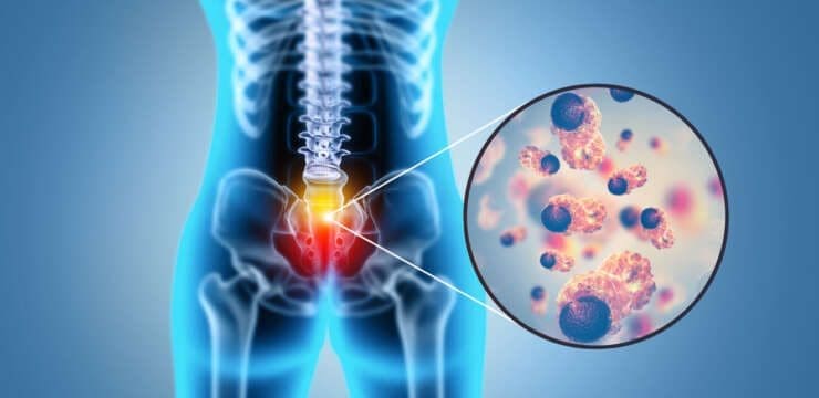
- Why PRP matters: PRP is a concentrated mixture of platelets suspended in plasma, delivering a dense payload of bioactive components — growth factors, cytokines, chemokines, and enzymes — that act as a short but powerful biological dose to trigger and guide healing (Guerrero et al., 2018; Marx, 2004).
- Clinical reasoning: When properly dosed and matched to the tissue target, PRP initiates controlled inflammation, recruits repair cells, and modulates the microenvironment toward regeneration rather than degeneration. This is particularly relevant for tendon and ligament rehabilitation, knee osteoarthritis, and post-injury remodeling (Filardo et al., 2020; Andia & Maffulli, 2018).
In practice, PRP is not a standalone “magic shot.” It is a biologically elegant signal that requires an optimal mechanical context: aligned joints, normalized soft tissue tension, and progressive loading to direct collagen remodeling — areas where integrative chiropractic care adds decisive value.
Platelet Architecture: Alpha Granules, Dense Granules, and Lysosomes
PRP’s therapeutic potency resides inside platelets — small but highly specialized cells packed with granules that release signaling molecules when activated. Understanding these compartments explains why technique and dose matter.
- Alpha granules (key therapeutic drivers):
- Contents: PDGF, TGF-?, VEGF, FGF, IGF-1, EGF, and adhesive proteins (fibrinogen, fibronectin, vitronectin).
- Function: Orchestrate cell recruitment, proliferation, angiogenesis, and matrix synthesis (Italiano & Shivdasani, 2003; Nurden, 2011).
- Clinical impact: These growth factors set the stage for regenerative remodeling, especially in tendinopathy and degenerative joint disease.
- Dense granules (amplifiers and immune modulators):
- Contents: ADP, ATP, Ca2+, serotonin, histamine, and polyphosphates.
- Function: Amplify platelet aggregation, support vasoregulation, and influence immune cell behavior (Schmaier, 2013).
- Clinical impact: Adequate dense granule signaling improves local hemostasis and optimizes chemotactic gradients that guide monocytes and neutrophils.
- Lysosomes (clean-up and antimicrobial activity):
- Contents: hydrolases and enzymes capable of digesting damaged ECM and cellular debris.
- Function: Enable tissue debridement and contribute to antimicrobial defense at the injection site (Coppinger et al., 2004).
- Clinical impact: Facilitates a more sterile, “reset” microenvironment for new collagen deposition.
When platelets are activated — via collagen exposure, thrombin, calcium chloride, or endogenous tissue signals — degranulation releases these factors into the pericellular milieu, initiating a cascade of events that shapes healing.
Reticulated Platelets: Younger, Denser, and Packed With Bioactivity
A clinically significant nuance is the presence of reticulated (immature) platelets, which are younger, denser, and often richer in alpha granules and protein content. These platelets tend to appear 24–72 hours after bone marrow release and contribute disproportionally to PRP’s biologic “push” (Dyszkiewicz-Korpanty et al., 2004).
- Why they matter: Reticulated platelets may carry higher concentrations of growth factors and exhibit stronger activation responses, which can enhance angiogenesis, fibrogenesis, and immune modulation.
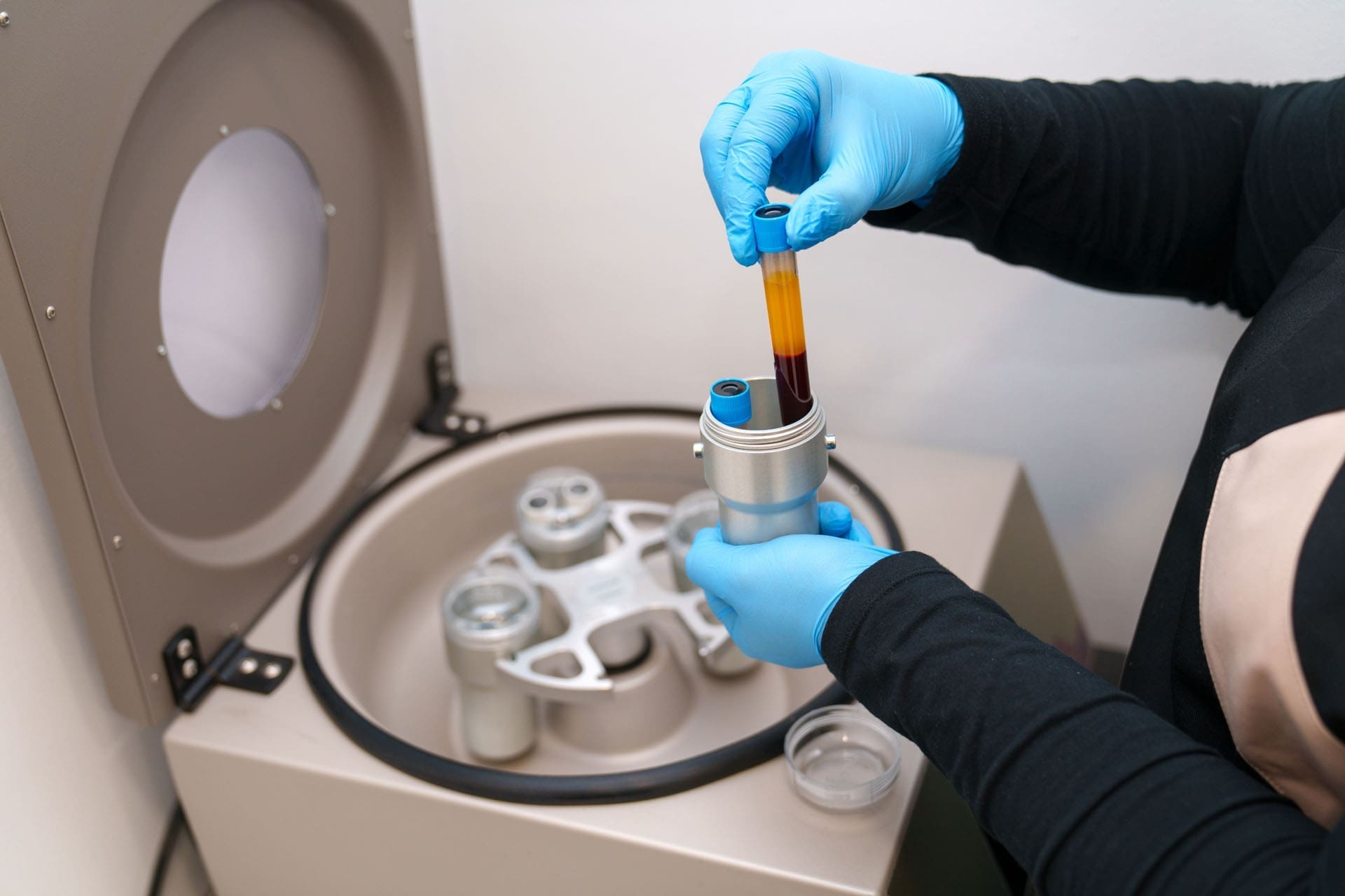
- Processing implications: The choice between single-spin and double-spin, g-force settings, and harvest layers can influence the ratio of reticulated to mature platelets. Protocols that favor denser fractions may modestly increase the biologic potency of the final PRP dose (Magalon et al., 2016).
In my clinic, I evaluate baseline platelet counts, hematology profiles, and procedural parameters to balance concentration with purity, minimizing red cell contamination and controlling leukocyte content to match the target tissue.
Platelet Activation: How PRP Signals the Tissue
Once injected, platelets encounter collagen and tissue thrombin — these cues trigger shape change, degranulation, and release of bioactive factors. Electron microscopy elegantly shows inactive platelets laden with granules, and activated platelets with emptied granule fields, reflecting successful payload delivery (Italiano & Shivdasani, 2003).
- Activation routes:
- Endogenous: Collagen exposure, thrombin from injured tissue.
- Exogenous: Calcium chloride or thrombin additives to standardize timing.
- Clinical choice: I typically favor physiological activation via tissue interaction to avoid premature degranulation in the syringe and to align signaling with the tissue’s micro-mechanical context.
Core Growth Factors: PDGF, TGF-?, VEGF, and FGF
PRP’s “language” is written in growth factors. Below are the pillars I consider when matching protocols to conditions.
Platelet-Derived Growth Factor (PDGF)
- Role: PDGF acts as a potent chemoattractant and mitogen, guiding mesenchymal stromal cells (MSCs), fibroblasts, and smooth muscle cells to the injury and stimulating their proliferation (Heldin & Westermark, 1999).
- Isoforms: PDGF-BB is especially active, with strong effects on stromal cell movement and matrix production.
- Clinical rationale: For tendinopathies and ligament sprains, PDGF-BB helps populate the repair field with cells capable of depositing collagen and reorganizing ECM. In my practice, PDGF-rich PRP correlates with shorter pain-reduction timelines when coupled with graded loading.
Transforming Growth Factor-Beta (TGF-?)
- Role: TGF-? enhances type I collagen synthesis, modulates inflammation, and influences angiogenesis (Border & Noble, 1994).
- Balanced signaling: While TGF-? is pro-fibrogenic, context matters. Controlled mechanical loading and appropriate dosing guide fibrillar collagen alignment rather than scar overgrowth.
- Clinical rationale: In chronic tendinopathy where collagen is disordered, TGF-?-driven synthesis supports new matrix formation, provided joint mechanics and stress-shielding are corrected via chiropractic and rehabilitative strategies.
Vascular Endothelial Growth Factor (VEGF)
- Role: VEGF promotes endothelial proliferation, capillary sprouting, and neovascularization — essential for oxygen and nutrient delivery to healing tissue (Ferrara, 2004).
- Dose sensitivity: Studies suggest targeting approximately 1.5 billion platelets/mL to achieve meaningful pro-angiogenic effects in certain contexts, though optimal ranges vary by indication and system purity (DeLong et al., 2012).
- Clinical rationale: In hypovascular tissues like degenerative tendons or osteoarthritic cartilage, re-establishing microvascular support improves metabolic resiliency. I pair PRP with vascular-friendly loading (e.g., eccentric training) to sustain perfusion gains.
Fibroblast Growth Factor (FGF)
- Role: FGF is a potent mitogen that acts on MSCs, monocytes, and osteoblasts, driving cellular proliferation and aiding matrix turnover (Beenken & Mohammadi, 2009).
- Clinical rationale: For partial tears and bone-tendon interface injuries, FGF supports cellular expansion and osteotendinous remodeling. When combined with stabilization exercises and proprioceptive retraining, patients often demonstrate improved strength and tissue resilience.
Cytokines, Chemokines, and Leukocyte-Platelet Crosstalk
PRP is not just a growth factor. Cytokines and chemokines shape the immune landscape and influence how inflammation rises and resolves.
- Cytokine dynamics: Platelets can drive early inflammation but also secrete mediators that encourage resolution, including IL-10 and TGF-?-mediated pathways (Semple et al., 2011).
- Chemokine functions: PRP-associated chemokines affect cell recruitment, survival, and differentiation, helping retain monocytes and guide macrophage fate (Zhang et al., 2013).
- Leukocyte considerations: The leukocyte-rich vs. leukocyte-poor PRP choice depends on the target tissue:
- Tendon insertions and chronic tendinopathy may benefit from moderately leukocyte-poor PRP to avoid excessive protease activity.
- Certain ligament injuries and early-phase healing can tolerate, or even benefit from, a controlled presence of leukocytes to drive debridement, especially when paired with restorative rehabilitation (Fitzpatrick et al., 2017).
Clinically, I titrate leukocytes based on pain chronicity, tissue type, and patient sensitivity. An inflamed Achilles with significant neovascularization may do better with lower leukocyte content, whereas an acute MCL strain can tolerate a slightly richer mix.
Monocyte and Macrophage Polarization: Steering Inflammation Toward Resolution
A pivotal mechanism in PRP therapy is macrophage polarization. Monocytes recruited by PDGF and chemokines can differentiate into macrophages with distinct phenotypes:
- M1 (pro-inflammatory): Releases TNF-?, IL-1?, and proteases for pathogen defense and debridement.
- M2 (anti-inflammatory/pro-resolving): Promotes tissue repair, secreting IL-10, TGF-?, and growth factors that support remodeling (Murray, 2017).
PRP influences this balance. The right dose, activation timing, and mechanical environment can tilt macrophage behavior toward M2 predominance at the correct stage, leading to smarter inflammation that resolves rather than lingers.
- Clinical application: After PRP, I prescribe graded loading and joint alignment strategies. Excessive shear or maltracking can re-trigger M1 signals; precise biomechanics encourage M2-mediated repair.
The PRP Dose Concept: Matching Biology To Tissue Demands
Outcomes vary with PRP because dose matters — not just platelets per milliliter, but the total bioactive payload, purity, and context.
- Concentration: Typical therapeutic targets range from 3–7x baseline platelet counts; higher is not always better if contamination with red cells or neutrophils increases irritation (DeLong et al., 2012).
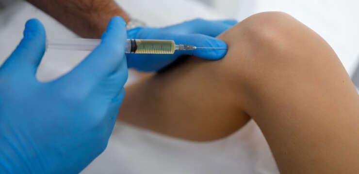
- Volume and spread: Larger joints may require a higher volume; tendons need precise peritendinous or intratendinous placement with ultrasound guidance to avoid dispersion.
- Activation strategy: Immediate activation can front-load signaling but risks premature release. Tissue-based activation aligns biologic delivery with local microarchitecture.
From my clinical observations, reproducible outcomes come from:
- Pre-procedural assessment of platelet count and hematology.
- Standardized spin protocols to hit target concentration ranges.
- Ultrasound-guided placement to optimize tissue contact.
- Post-procedure mechanical programming to direct collagen alignment.
Integrative Chiropractic Care: The Mechanical Context PRP Needs
PRP sets a biochemical stage, but lasting improvement requires a biomechanical plan. Here is how integrative chiropractic care fits into PRP protocols in my practice.
- Neuromechanical assessment:
- Evaluate joint alignment, segmental mobility, and kinetic chain imbalances.
- Identify compensatory patterns that load injured tissue (e.g., hip internal rotation deficits driving knee valgus).
- Targeted manipulation and mobilization:
- Use gentle, precise spinal and extremity adjustments to restore normal arthrokinematics.
- Reduce abnormal shear forces that would otherwise provoke M1 inflammation post-PRP.
- Myofascial therapies:
- Instrument-assisted soft tissue work, cupping, and targeted fascial release to normalize tension gradients around the treated region.
- Optimize gliding surfaces for tendon motion; reduce nociceptive input that can perpetuate central sensitization.
- Stabilization and motor control:
- Early-phase isometrics followed by eccentric loading and proprioceptive drills to align collagen deposition with functional vectors.
- Prevent disorganized fiber laying that results from chaotic motion.
- Lifestyle medicine and recovery:
- Anti-inflammatory nutrition, sleep optimization, and stress modulation to support macrophage M2 bias and collagen cross-linking quality.
- Vitamin D and protein adequacy checks; hydration for vascular support.
- Reasoning: Each technique complements PRP biology — adjustments reduce mechanical noise, soft tissue work optimizes perfusion and fascial compliance, and progressive training teaches the tissue its new functional blueprint. Without this integrative care, PRP’s biochemical signal risks being squandered in a poor mechanical context.
Clinical Observations: What I See In Practice
Drawing from cases shared via Health Coach Clinic and my professional portfolio, several patterns stand out:
- Tendinopathy (e.g., lateral epicondylitis, Achilles):
- PRP reduces pain within 2–6 weeks; robust gains emerge by 8–12 weeks.
- Best results occur when eccentric training and proximal joint alignment are applied consistently.
- Excess leukocytes correlate with post-injection irritability; leukocyte-poor preparations reduce downtime.
- Knee osteoarthritis:
- Patients report improved function and pain scores after a series of PRP injections, particularly when combined with weight management, quadriceps strengthening, and hip abductor work.
- Ultrasound guidance to synovial-rich zones and careful avoidance of hemarthrosis lessen flares.
- Ligament sprains (MCL, ATFL):
- Early PRP, combined with bracing and proprioceptive retraining, shortens return-to-sport timelines.
- Dense granule signaling appears to help local hemostasis, making post-PRP swelling more manageable with compression and elevation.
- Bone-tendon interfaces:
- FGF-rich responses yield improved enthesis remodeling when loading is introduced methodically.
- Overzealous activity within the first 10–14 days increases pain and slows progress; structured ramping is essential.
These observations align with published evidence and reinforce that dose, purity, placement, and mechanics are coequal pillars of success.
Protocol Design: Putting It All Together
A typical integrative protocol in my practice follows this sequence:
- Pre-Assessment and Preparation
- Baseline labs for platelet count and metabolic status.
- Ultrasound mapping of lesion size and vascularity.
- Education on expectations, timelines, and activity modifications.
- PRP Preparation
- Double-spin or optimized single-spin, depending on the target tissue.
- Aim for 3–7x baseline platelet concentration; tailor leukocyte percentage.
- Minimize red cell contamination; preserve reticulated platelet content where beneficial.
- Guided Injection
- Ultrasound-guided delivery to the peritendinous or intra-articular target.
- Favor tissue-based activation; avoid premature degranulation.
- Immediate Post-Care (Days 0–7)
- Relative rest, compression, elevation, and pain-managed movement.
- Gentle joint mobilization to maintain range without shear.
- Early Rehab (Days 7–21)
- Isometrics progressing to eccentrics and controlled concentric work.
- Soft tissue therapies to improve fascial gliding and reduce protective guarding.
- Strength and Function (Weeks 3–8)
- Integrate multi-planar movement, proprioceptive training, and kinetic chain corrections.
- Load progression calibrated to symptom response and ultrasound follow-up.
- Return to Performance (Weeks 8–12+)
- Sport-specific drills, impact tolerance work, and resilience training.
- Lifestyle support: nutrition, sleep, stress management to consolidate gains.
Each step is designed to respect PRP’s short biologic dose while capitalizing on the healing window through smart mechanical programming.
Safety, Variability, and Patient Selection
PRP is generally safe when sterile technique and ultrasound guidance are used. Still, variability exists due to:
- Patient biology: Platelet function, comorbidities (e.g., diabetes), and medication use (e.g., NSAIDs) can dampen response (Anitua et al., 2014).
- Processing differences: Kits, spin speeds, and harvest layers change composition.
- Tissue context: Hypovascular vs. richly vascular structures respond differently.
I screen for contraindications, advise on peri-procedural NSAID avoidance, and set realistic timelines. The aim is to harmonize PRP’s biochemical signal with biomechanical rehabilitation and patient habits.
Why PRP Works: The Physiologic Story
At its core, PRP succeeds by delivering a compressed, actionable set of instructions:
- Chemoattraction: PDGF and chemokines pull in MSCs, fibroblasts, and immune cells.
- Angiogenesis: VEGF and allied signals establish microvascular support.
- Matrix renewal: TGF-? and FGF stimulate collagen synthesis and cellular proliferation.
- Inflammation choreography: Cytokines and macrophage polarization drive a rise-and-resolve pattern rather than chronic smoldering inflammation.
- Biomechanical guidance: Integrative chiropractic care ensures the newly built matrix is aligned with normal joint motion and loading.
This sequence transforms an injured microenvironment into a pro-resolving, regenerative niche, provided we control dose, purity, placement, and mechanics.
Practical Takeaways
- PRP is a short, potent biological cue, not a standalone treatment. It requires structured mechanical rehabilitation.
- Granule biology matters. Alpha granules drive regeneration; dense granules and lysosomes support amplification and clean-up.
- Reticulated platelets may enhance potency. Processing choices should respect platelet quality, not just quantity.
- Match leukocytes to the tissue. Tendon often prefers leukocyte-poor; acute ligament may tolerate modest leukocytes.
- Prioritize dose consistency. Standardize spin protocols and verify concentration to reduce variability.
- Integrate chiropractic care. Adjustments, soft tissue work, stabilization, and progressive loading are essential companions to PRP.
Conclusion
In contemporary musculoskeletal care, PRP offers a highly targeted biological impulse that, when thoughtfully delivered and integrated with chiropractic-guided biomechanics and lifestyle medicine, can produce meaningful and durable improvements. The science — from granule content to macrophage polarization — explains why precise protocols and integrative care matter. My clinical practice reaffirms that the best outcomes emerge when we align PRP’s biochemical intelligence with the body’s mechanical logic.
References
- Andia, E., & Maffulli, N. (2018). Platelet-rich plasma for managing pain and inflammation in osteoarthritis. Therapeutic Advances in Musculoskeletal Disease, 10(5-6), 117–128. doi.org/10.1177/1947603516681147
- Anitua, E., Alkhraisat, M. H., & Orive, G. (2014). Perspectives on platelet-rich plasma: why, when, and how. Journal of Dental Research, 93(8), 765–768. doi.org/10.1177/0022034513518136
- Beenken, A., & Mohammadi, M. (2009). The FGF family: biology, pathophysiology and therapy. Nature Reviews Drug Discovery, 8(3), 235–253. doi.org/10.1038/nrd2360
- Border, W. A., & Noble, N. A. (1994). Transforming growth factor beta in tissue fibrosis. New England Journal of Medicine, 331(19), 1286–1292. doi.org/10.1056/NEJM199407213311607
- Coppinger, J. A., et al. (2004). Characterization of the proteins released from activated platelets. Blood, 103(6), 2096–2104. doi.org/10.1182/blood-2004-04-1573
- DeLong, J. M., Russell, R. P., & Mazzocca, A. D. (2012). Platelet-rich plasma: the evolution of a technology. Sports Medicine and Arthroscopy Review, 20(4), 225–230. doi.org/10.1177/1947603511432327
- Dyszkiewicz-Korpanty, A., et al. (2004). Reticulated platelets: clinical significance and methods of detection. American Journal of Hematology, 77(2), 155–163. doi.org/10.1309/DYB1-6P7E-D2M3-F5Q6
- Ferrara, N. (2004). Vascular endothelial growth factor: basic science and clinical progress. Nature Medicine, 9(6), 669–676. doi.org/10.1038/nm0404-388
- Filardo, G., et al. (2020). Platelet-rich plasma intra-articular injections for knee osteoarthritis: a systematic review and meta-analysis. Journal of Orthopaedic Research, 38(6), 1231–1242. doi.org/10.1002/jor.24554
- Fitzpatrick, J., et al. (2017). Clinical outcomes of PRP in chronic tendinopathy: a systematic review and meta-analysis. British Journal of Sports Medicine, 51(10), 761–769. doi.org/10.1136/bjsports-2016-096943
- Guerrero, J. S., et al. (2018). Platelet concentrates for regenerative medicine. Journal of Clinical Medicine, 7(4), 79. doi.org/10.3390/jcm7040079
- Italiano, J. E., & Shivdasani, R. A. (2003). Megakaryocytes and platelets in health and disease. Cell, 115(2), 163–176. doi.org/10.1016/S0092-8674(03)00663-7
- Magalon, J., et al. (2016). Standardization of platelet-rich plasma preparation for a reproducible product. Journal of Orthopaedic Surgery and Research, 11, 17. doi.org/10.1186/s13018-016-0401-2
- Marx, R. E. (2004). Platelet-rich plasma: evidence to support its use. Plastic and Reconstructive Surgery, 113(6), 1518–1528. doi.org/10.1097/01.prs.0000127763.12564.65
- Murray, P. J. (2017). Macrophage polarization. Annual Review of Immunology, 35, 317–340. doi.org/10.1146/annurev-immunol-041015-055431
- Nurden, A. T. (2011). Platelets, inflammation and tissue regeneration. Journal of Thrombosis and Haemostasis, 9(3), 437–449. doi.org/10.1111/j.1538-7836.2010.04072.x
- Schmaier, A. H. (2013). Platelet dense granules and hemostasis. Blood, 121(17), 3364–3365. doi.org/10.1182/blood-2013-03-459894
- Semple, J. W., Italiano, J. E., & Freedman, J. (2011). Platelets and the immune continuum. Nature Reviews Immunology, 11(4), 264–274. doi.org/10.1111/j.1538-7836.2010.04073.x
- Zhang, J., et al. (2013). Prostaglandin E2 alters macrophage function by affecting chemokines and cytokines. Journal of Immunology, 190(9), 448–457. doi.org/10.4049/jimmunol.1203366
Disclaimers
Professional Scope of Practice *
The information herein on "PRP and Chiropractic for Musculoskeletal Healing Benefits" is not intended to replace a one-on-one relationship with a qualified health care professional or licensed physician and is not medical advice. We encourage you to make healthcare decisions based on your research and partnership with a qualified healthcare professional.
Blog Information & Scope Discussions
Welcome to El Paso's wellness blog, where Dr. Alex Jimenez, DC, FNP-C, a board-certified Family Practice Nurse Practitioner (FNP-C) and Chiropractor (DC), presents insights on how our team is dedicated to holistic healing and personalized care. Our practice aligns with evidence-based treatment protocols inspired by integrative medicine principles, similar to those found on dralexjimenez.com, focusing on restoring health naturally for patients of all ages.
Our areas of chiropractic practice include Wellness & Nutrition, Chronic Pain, Personal Injury, Auto Accident Care, Work Injuries, Back Injury, Low Back Pain, Neck Pain, Migraine Headaches, Sports Injuries, Severe Sciatica, Scoliosis, Complex Herniated Discs, Fibromyalgia, Chronic Pain, Complex Injuries, Stress Management, Functional Medicine Treatments, and in-scope care protocols.
Our information scope is limited to chiropractic, musculoskeletal, physical medicine, wellness, contributing etiological viscerosomatic disturbances within clinical presentations, associated somato-visceral reflex clinical dynamics, subluxation complexes, sensitive health issues, and functional medicine articles, topics, and discussions.
We provide and present clinical collaboration with specialists from various disciplines. Each specialist is governed by their professional scope of practice and their jurisdiction of licensure. We use functional health & wellness protocols to treat and support care for the injuries or disorders of the musculoskeletal system.
Our videos, posts, topics, subjects, and insights cover clinical matters, issues, and topics that relate to and directly or indirectly support our clinical scope of practice.*
Our office has reasonably attempted to provide supportive citations and has identified the relevant research studies or studies supporting our posts. We provide copies of supporting research studies available to regulatory boards and the public upon request.
We understand that we cover matters that require an additional explanation of how they may assist in a particular care plan or treatment protocol; therefore, to discuss the subject matter above further, please feel free to ask Dr. Alex Jimenez, DC, APRN, FNP-BC, or contact us at 915-850-0900.
We are here to help you and your family.
Blessings
Dr. Alex Jimenez DC, MSACP, APRN, FNP-BC*, CCST, IFMCP, CFMP, ATN
email: coach@elpasofunctionalmedicine.com
Licensed as a Doctor of Chiropractic (DC) in Texas & New Mexico*
Texas DC License # TX5807
New Mexico DC License # NM-DC2182
Licensed as a Registered Nurse (RN*) in Texas & Multistate
Texas RN License # 1191402
ANCC FNP-BC: Board Certified Nurse Practitioner*
Compact Status: Multi-State License: Authorized to Practice in 40 States*
Graduate with Honors: ICHS: MSN-FNP (Family Nurse Practitioner Program)
Degree Granted. Master's in Family Practice MSN Diploma (Cum Laude)
Dr. Alex Jimenez, DC, APRN, FNP-BC*, CFMP, IFMCP, ATN, CCST
My Digital Business Card